Share   Subscribe to RSS feed

Brian Schiff’s Blog

Injury Prevention, Sports Rehab & Performance Training Expert

Well, after a silent stretch the past 2 weeks or so related to my study/preparation for the OCS exam, I am back to blogging! Today’s post is a pertinent one for runners and athletes suffering from lower limb injuries.

Static stretching has taken a bit of a beating in the strength and conditioning world in the last few years. Dynamic warm-ups and active mobility have taken center stage as of late.  While these active modalities are certainly superior for prior to practice, play and ballistic activity periods, I still believe stretching has a place in rehab and conditioning.

caf-step-stretch

Interestingly enough, a study recently published in the March 2012 Journal of Strength & Conditioning Research examined the effects of static stretching of the calf and its impact on the strength/ROM of the contralateral side.  Click here for the abstract.

image_thumb60

In a nutshell, the authors had two groups of untrained individuals: test group (6 male and 7 female subjects) and control group (6 male and 6 female subjects) who participated.  The test group did supervised active right calf stretching 3 days per week for 10 weeks (four 30 sec stretches w/30 sec of rest between stretches).  They stood on a beam 30 cm above the floor with the left knee slightly bent to offload the left leg as well as placing the hands on the wall while they leaned forward allowing the right heel to drop toward the floor until a max tolerable stretch was felt.  The knee was straight throughout on the stretch side.

Control subjects did no stretching at all.  All subjects were instructed to maintain their normal physical activity but refrain from any resistance training or stretching during the 10 week investigation.  The results:

  • 29% increase in 1RM calf raise strength on the right
  • 11% increase in 1RM calf raise strength on the left
  • significant 8% increase in calf ROM on right
  • significant 1% decrease in calf ROM on the left (non stretch side)
  • No change in strength or ROM for control group

The authors conclude that the results of this study best apply to rehab settings.  For example, they suggest that this procedure may be an effective way to combat the loss of strength in limbs that have been immobilized after injury or surgery simply by stretching the mobile (unaffected) side.  They also point out that this may be a way to mitigate strength loss when access to traditional strengthening modalities are not readily available.

Clearly, athletes suffering an acute ankle sprain as well as runners suffering soleus/Achilles/lower limb overuse injuries would benefit from such a strategy. So why does this work?  Zhou in earlier work describes a cross training effect due to neural adaptations regulated in the spinal cord.

What does this mean for you and me?  Well, as someone who works with many runners I am always looking at eccentric control of the G/S complex as well as effective single leg heel raise strength.  The idea that stretching the uninvolved side to strengthen the involved side seems like a no brainer.  Clients suffering from tendonitis, plantar fasciitis, stress reactions, sprains and other injuries can use this as an early intervention without stressing the involved side.

More importantly, I like the idea of increasing neural adaptation and ROM in the stretch side through eccentric load as the dissipation of ground reaction forces will be more efficient in a calf that effectively handles eccentric loads through a sufficient range of motion.  This study definitely highlights the importance of stretching in novice runners and those with tight gastrocs. I am curious if the bent knee stretch would have had a similar effect primarily on ROM – perhaps they will investigate that further in the future.

As we move more toward mid and forefoot running gait, I believe the fitness of the G/S complex will be even more important than before as stress is transferred away from the knee and more toward the foot/ankle complex.  Clearly, we need more studies in trained subjects on unilateral stretching to determine if the same effects and degree of impact will be seen, but this study shows some promise for active static calf stretching in the appropriate populations.

It is widely accepted that decreased hip strength and stability leads to knee valgus. Excessive frontal plane motion and valgus torque increase the risk for non-contact ACL injuries. While we know that hip abductor weakness is more of an issue in females than males, the question remains to what degree other factors are involved.

knee-valgus-add-ir

Claiborne et al (1) noted that only 22% of knee variability could be linked solely to hip muscles surrounding the hip. In light of this we must look at the whole kinetic chain when assessing movement dysfunction and injury risk. In the most recent issue of the IJSPT, researchers sought to discover how activating the core during a single leg squat would impact the kinematics of 14 female college-age women. They excluded participants with current pain or injury to the lower extremities or torso, or if they had a history of any lower extremity injuries or surgeries in the past 12 months.

The participants were assessed for their capacity to recruit core stabilizer muscles using lower abdominal strength assessment as described by Sahrman (2). This testing model has 5 levels of increasing difficulty used to challenge participants to maintain a neutral spine. The draw back of this method is that it is done in supine versus the standing position of this study, but the author acknowledges this limitation. Out of a possible high score of 5, five of the participants scored a 1 or 2, while the other nine scored a zero.

For the study, a six inch step was used to assess 2 reps of a single leg squat. Each participant was asked to perform the test with the dominant leg to standardize conditions. They performed the reps under two conditions:

1. CORE – engaged abdominal muscles as they had been instructed to do so during the Sahrman test

2. NOCORE – no purposeful engagement of abdominal muscles

Results

  • The CORE condition resulted in smaller hip displacements in the frontal plane but had no effect on hip angular range of motion – essentially there was less medial/lateral movement
  • The CORE group did not exhibit any changes in knee displacement but did exhibit greater degree of knee flexion – this may suggest higher function assuming more knee flexion is desired during squat tasks in sports and functional activities
  • Those scoring the lowest on the core assessments had larger improvements in performance when they did in fact activate the core musculature

How do we use this information to affect our practice?  Well, in terms of rehab it seems straightforward and many of us may already encourage patients to activate their core during treatment.  However, I think the greater contribution may come in injury prevention programs (particularly ACL programs) where we are looking at all facets of neuromuscular control and appropriate muscle activation patterns.

With any prehab or rehab strategy, we as clinicians, trainers and strength coaches are essentially trying to reprogram the brain to summon and execute a better motor pattern or strategy – feedforward training.  We know that healthy individuals tend to have better transverse abdominus and multifidus muscle activation, so it only makes sense to consider activation of local stabilizers as we work on global muscles.  Improving core and pelvic stability should only help reduce unwanted frontal plane motion.

With that said, the authors of this study readily acknowledge more work needs to be done with larger clinical populations (including EMG work) to more clearly identify what magnitude the core musculature has on lower extremity motion and displacement.

Keep in mind the proper program will always stem from your ability to assess movement impairment and tissue dysfunction.  I suggest beginning with a FMS in the athletic population and incorporating parts of that or the SFMA to compliment your evaluation in the clinic.  This will generally reveal the priorities for the corrective exercises.  For now, we can use this information in this particular study to be more intentional with our patients and clients suffering knee and hip dysfunction by adding this one simple step to our programming.

References

1. Claiborne TL, Armstrong CW, Gandhi V, Princivero DM. Relationship between hip and knee strength and knee valgus during a single leg squat. Journal of applied biomechanics. 2006;22(1):41.

2. Faries MD, Greenwood M. Core training: stabilizing the confusion. Strength & Conditioning Journal. 2007 ;29(2):10.

Dysfunctional movement is common with shoulder pain and impingement.  One dysfunction you may encounter is a downwardly rotated scapula. If upward rotation is limited, a client will display excessive shoulder flexion above 90 degrees when the humerus is in maximal internal rotation.  Typically, a person will have minimal flexion beyond 90 degrees if the scapula is moving properly.

Upward rotation of the scapula is the result of a force couple between the upper and lower trap along with the serratus anterior.  If any of these muscles are weak, rotation can be limited and overpowered by the rhomboids and levator scapulae muscles (both downward rotators).  This pattern of muscle dominance is common.

Additionally, tightness in the rhomboids, levator scapulae, pec minor or latissimus can also restrict normal mobility.  It is probably safe to assume stretching of the chest and lats would be helpful, but it is critical to encourage the proper muscle firing patterns in the traps and serratus anterior as well.

Below is a video demonstrating wall slide shrugs.  The shrug should be done at or above 90 degrees.  You can perform reps at multiple angles or move to end range and perform a series there.

Application:  The exercise is designed to encourage upward rotation in a more functional manner as opposed to traditional shrugs with the arms at the side.  While I am not opposed to traditional shrugs with little or no weight for basic elevation, this position generally tends to activate the rhomboids and levator scapulae which is not desired given their natural dominance pattern.

The wall slide shrugs should not create any pain or discomfort.  However, they may feel awkward particularly if the client has a faulty muscle activation pattern.  As muscle tightness resolves and strength improves, clients should gain more mobility and optimal shoulder function.

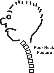
It is no secret that all the technology in our world has made poor posture even more prevalent than before. I always say the brain picks the path of least resistance for the body and that tendency combined with gravity leads to a natural forward head posture. Hours spent on the computer, texting and sitting often promote a picture like this:

poor-neck

This positioning leads to lower cervical flexion and upper cervical extension thereby creating muscle imbalances.  Keep in mind the rest of the spine takes its cues from the head and neck.  A forward head encourages thoracic kyphosis, shoulder internal rotation and lumbar flexion.  The back muscles give way to hanging on the static structures and the brain grows accustomed to this relaxed posture.  The result = increased strain, muscle tension and mechanical deformation.  In addition, neuromuscular activation of the proper postural muscles is compromised or shut down completely.

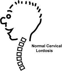
This sequence of events often gives way to postural pain, tightness, tension headaches and eventually even more intense pain and symptoms related to disc pathology.  Essentially, the wrong structures spend too much time under tension, while our muscles go on a prolonged siesta throughout the day.  Good posture takes hard work and looks like this:

normal-neck

So, some simple strategies to improve posture include an awareness of proper alignment (ears in line with the shoulders), a conscious effort to sit and stand with better posture and consistent postural strengthening.  One of my favorite exercises to do is a simple prone neck retraction (see video below)

For a complete description of the exercise, click here to read my full column in PFP Magazine.

Finally, I want to leave you with some information from another clinician, Joe Brence, DPT on pain classification. He addresses the biomedical versus biopsychosocial approach to treating pain on The Manual Therapist’s blog.  I tend to believe we must use components of both systems as well as our intuition and clinical experience, but it is an informative post.  Click here to read his guest blog post on this.

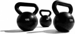
six-pack-shortcuts-kettlebell-cardio-routines

Kettlebells are very popular training tools these days.  I find them useful in many ways – improving grip strength, core activation, asymmetrical loading, etc.  With that said, I also feel with movement flaws and/or improper technique, they carry an inherent injury risk.

It is interesting to note that some people find swings to be very therapeutic and good for their back, while others who are capable of lifting very high loads with traditional lifts find them to be irritating to the spine.  So why is this?

If you are like me, knowing the “why” or “cause and effect” behind exercise is very important.  I am not one to blindly use an exercise without knowing its intended purpose and then quantifying risk vs. reward and results. So, it was with great interest I read Stuart McGill and Leigh Marshall’s recent article on kettlebell swings, snatches and bottoms-up carries in the NSCA Journal of Strength & Conditioning Research (Jan 2012).

Click here for the abstract.

While the sample size is small, I think the article provides some gems in regard to training given no one has really looked at spine loading during various swings and carries.  The authors used surface EMG to record muscle activation of the back, hip and core muscles throughout the various exercises – swing, swing with Kime (abdominal pulse at top of the swing), swing to snatch, racked carry and bottoms-up carry.

Without going into all the tiny details, I wanted to share what I consider to be some key takeaways for rehab and training:

  • Unlike traditional low back extension exercises such as lifting a bar or squatting exercises, the swing creates a relatively high posterior shear force (namely L4 on L5) in relation to the compressive load – this may explain why some powerlifters have no issues with heavy dead lifts but are bothered by swings
  • Both compressive and shear forces were highest at the beginning of the swing
  • From a compressive standpoint loads with a 16 kg kettlebell (swing) are less than one-half of that of lifting 27 kg on an Olympic bar and these would seemingly pose a low relative injury risk
  • KB swings do appear to require sufficient spine stability in this shear mode to ensure that is is a “safe” exercise selection
  • Those with back pain develop movement flaws and the authors report one of the most common is to move the spine under load instead of the hips – so instead of hip hinging, injured clientele are more apt to shift or bend the spine leading to repetitive and harmful forces
  • A modified approach to swings with careful cueing and progression is suggested for clinicians
  • The bottoms-up carry poses more challenge to the core musculature likely due to requiring more grip strength (thus stiffening the core per McGill in Ultimate Back Fitness & Performance) as well as necessitating more control to carry it, hence making it a good choice for training in terms of activation of these muscles

So, in my mind kettlebell training (like any other form of training) requires proper form, movement assessment and an intimate knowledge of the client’s medical and training history.  In addition to that, we must carefully scrutinize execution of the exercise and deliver appropriate feedback and analysis.

While maximal shear occurs at the bottom, I cannot help but wonder about the potential impact of tight iliopsoas muscles given their unique relationship to the lumbar spine and reverse muscle action.  It would be interesting to know if those with a greater anterior tilt and tightness are more likely to experience higher shear forces or potential back soreness over time.

This brings the discussion back to quality of movement and movement assessment.  In my mind, adequately assessing the hips (flexibility, strength and stability) is also a key variable in determining how best to approach integrating the swings.  As Gray would say, the lumbar spine needs stability while the hips require mobility.

A lack of hip mobility is definitely a relative precaution for swings in my mind.  On top of that, fundamental hip strength/stability and core strength should be evident.  Perhaps even regressing to rudimentary hip thrusts and bridges may be the best place to start for those needing a primer on form and proper movement before moving to a basic swing.

Nonetheless, a big thanks to Stuart McGill and Leigh Marshall for this work and giving us some practical food for thought.  I hope this information helps you as much as it did me.  May your training be safe and effective!