Share   Subscribe to RSS feed

Brian Schiff’s Blog

Injury Prevention, Sports Rehab & Performance Training Expert

Femoroacetabular impingement (FAI) is often a hidden and misunderstood cause of hip pain.  I currently work with a physician who has studied under some of the best hip arthroscopists in the US, and he is performing arthroscopic procedures to resolve hip impingement.  For many years, this has likely been a source of misdiagnosed, under treated and debilitating hip pain for people.

As things advance in medicine, hip arthroscopy is expanding and allowing for easier surgical correction of these issues. However, it is not an easy surgery technically speaking.  As such, finding the right surgeon (if needed) is critical to attaining a positive outcome.  Who normally gets it?  Unfortunately, many people are predisposed to it, much like we see the natural genetic architecture (shape) of the acromion affecting impingement in the shoulder.

If you have an overhang of the hip acetabulum (socket) or non-spherical shape of the femoral head (or both) this can compromise the joint space and injure the joint cartilage and/or labrum.  Destruction can occur at a very young age.  I am currently rehabbing a 19 y/o male who recently underwent hip arthroscopy to debride his labrum and smooth out the hip socket and re-shape the femoral head.  He had extensive damage at an early age due to his joint architecture and shows some signs of impingement on the other side as well.

How do you know if you have hip impingement?  Generally, you may have hip joint pain along the front, side or back of the hip along with stiffness or a marked loss of motion (namely internal rotation).  It is common in high level athletes and active individuals.  However, other things may cause hip pain as well such as iliopsoas tendonitis, low back pain, SI joint pain, groin strain, hip dysplasia, etc. so a careful history, exam and plain films are necessary to confirm the diagnosis.  If suspected, an MR athrogram is usually ordered to confirm if there are labral tears present.  Physicians also use an injection with anesthetic to see if the pain is truly coming from the hip joint.  This may be done under fluoroscopy to ensure it is in the joint space.

Signs and symptoms of FAI may include:

  • Pain with sitting
  • Pain or limited squatting
  • Stiffness and decreased internal rotation
  • Pain with impingement testing (see picture below of hip flexion, adduction and internal rotation – examiner will move the hip into this position and marked stiffness/loss of internal rotation and pain indicates a positive test)

hip-impingement-test

Conservative treatment typically involves limiting or avoiding squats, strengthening the core and hip stabilizers as well as attempting to maximize mobility of the joint.  Due to the fact that by the time pain brings patients in to see the doctor there has already been marked labral and joint damage, a cautious and proactive approach to managing hip pain is warranted especially in younger active patients and athletes.

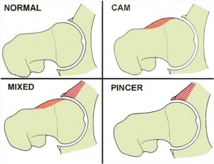
The types of lesions seen are either Cam or Pincer lesions.

Cam lesion – involves an aspherical shape of the femoral that causes abnormal contact between the ball and socket leading to impingement

Pincer lesion – involves excessive overgrowth of the acetabulum resulting in too much coverage of the femoral head and causing impingement where the labrum gets pinched

You can also see a mixed lesion where Cam and Pincer lesions are involved.  FAI may lead or contribute to cartilage damage, labral tears, hyperlaxity, sports hernias, low back pain and early arthritis.
fais

The good news is that these patients typically do well post-operatively.  Dr. Philipon et al reported in 2007 in the Knee Surg Sports Traumatol Arthrosc. (click here to read the abstract) on 45 professional athletes who underwent arthroscopic management of FAI with an average follow-up of 1.6 years.   In this time period 78% of them were able to return to their sport.

Following surgery, weight bearing may be restricted for the first 4 weeks or so to protect the labrum if it is repaired.  With a simple debridement and re-contouring of the acetabulum, weight bearing may be initiated earlier.  Avoiding twisting motions and excessive external rotation is a must in the first month or so as well.  Typically, impact and twisting restrictions are lifted around 3 months post-op.

In the end, proper diagnosis and treatment is necessary to preserve the hip joint and maximize function and return to sport.  If you or someone you know suffers from chronic and persistent hip pain that has failed conservative treatment, then consider getting a second look to rule out FAI.

By far the most comments on my blog and emails that flood my inbox these days have to do with SLAP tears.  I must admit that outside of ACL tears and rotator cuff issues, I find myself increasingly drawn to studying and researching this issue.  It definitely is a source of great pain for many and an issue that medical professionals are challenged by today.

In my personal clinical experience, I see good, bad and in between outcomes.  Through email and my blog I tend to read more on the not so good side from people who are seeking my expertise in how to resolve their issues.  When I speak to surgeons, I find they are often hesitant to commit to a set algorithm of treatment, and they are not 100% sure what the right answer is in addressing these injuries as a whole.

If you read the literature, the success in terms of patient satisfaction and return to premorbid activity levels is not going to make you rush down to the operating room and opt for an arthroscopic repair if you are an overhead athlete (especially baseball players).  However, other studies have presented more favorable data ranging from 63%-75% good-excellent satisfaction in other overhead athletes who have had the procedure done.

If you are unfamiliar with SLAP tears, I suggest reading my original post on them (click here).  In today’s post, I wanted to present a quick recap on Type II SLAP tears and some new published research on the results of revision procedures where the primary repair failed.

Below are two images of a type II tear (MRI and operative view from the scope)

type-2-slap-mri

type2slap

Keep in mind a type II tear means the biceps anchor/superior labrum has pulled away from the glenoid with resulting instability of the complex.  This is the most common type of tear seen among injured people.  In a study from the Kerlan-Jobe Orthopaedic Clinic in LA in the latest American Journal of Sports Medicine (June 2011 – click here for the abstract), they discussed a chart review of from 2003-2009 looking at patients who had undergone revision type II SLAP repairs.


Continue reading…

ACL injuries continue to proliferate among female athletes.  I am passionate about preventing them, and part of my professional mission is to study and evolve in my rehab and prevention training approaches all the time to stay on top.  I wanted to pass along some new information on a new screening tool just unveiled in the Strength and Conditioning Journal this month.

Before I reveal the screening and training tool, I want to take a moment and review what Timothy Hewett refers to as modifiable risk factors that contribute to injury risk based on his work:

  1. Ligament Dominance – defined as an imbalance b/w neuromuscular and ligamentous control  of dynamic knee stability and it is visualized by loss of frontal plane control with landing and cutting
  2. Quadriceps Dominance – defined as an imbalance between quad and hamstring strength, recruitment and coordination
  3. Leg Dominance – defined as an imbalance between the two legs with respect to strength, coordination and control
  4. Trunk Dominance ‘Core’ Dysfunction – defined as an imbalance b/w the inertial demands on the trunk and its ability to resist or control/resist it

Previously, Hewett has identified that high knee abduction moments are related to high LOAD on the knee and a major risk factor for ACL injury.  He and his colleagues have done extensive motion analysis in their lab in Cincinnati, OH.  As such, a drop landing test has been used as one tool to observe landing mechanics and assign some risk value to athletes competing in cutting and jumping sports.

In the current article (click here for the abstract) Meyer, Brent, Ford and Hewett unveil a new screening tool involving the tuck jump.  They propose that this tool is easier for the S & C coaches to do on the field and not only assess risk factors by way of observing technical flaws, but also use the tool as a training maneuver.

The idea is the subject will perform tuck jumps for 10 seconds consecutively while the observer makes notes on the following pre, mid and post jumping:

  1. Lower extremity valgus at landing
  2. Thighs do not reach parallel (peak height of jump)
  3. Thighs not equal side-to-side (during flight)
  4. Foot placement not shoulder width apart
  5. Foot placement not parallel (front to back)
  6. Foot contact timing not equal
  7. Excessive landing contact noise
  8. Pause b/w jumps
  9. Technique declines prior to 10 seconds
  10. Does not land in same footprint (excessive in flight motion)

Factors 1-3 refer to knee and thigh motion, 4-7 refer to foot position during landing and 8-10 refer to plyometric technique.  Coaches are instructed to grade the flaws if seen with check marks during the phases they are seen and use this as a guide for correction.  They may also use cameras in the frontal and sagittal plane to assist them.

My thoughts on this are:

  • There is sound science behind the rationale for the test and modifiable risk factors
  • There is a need for basic no-cost screening tools coaches can apply in their settings
  • The tuck jump assessment will provide instant feedback on form and identify technical flaws that may indicate higher risk for injury
  • The tuck jump is a higher demand plyo drill so I fear poor form may be as much to blame on inexperience and unrefined motor patterns as it is to just dominance patterns so we need to keep plyo training experience in mind when analyzing the screen results especially for beginners
  • The tuck jump assessment does not really consider fundamental movement restrictions that may bias the form on one side if an asymmetry is present
  • I still wonder how much ankle pronation impacts landing and whether we will see more research on this – there was a study done at ECU where they used orthotics and saw a reduction in ACL tears in their collegiate athletes so I have to wonder about this crucial element of the kinetic chain

In the end, we still lack many answers.  According to data published in the Journal of Athletic Training in 2006, non targeted neuromuscular training programs need to be applied to 89 female athletes to prevent 1 ACL tear.  So, we need to keep studying and applying science to our training, all the while critically questioning science and looking at our athletes holistically to find the best prevention strategies for each one individually and for at risk athletes as a whole.

I have been attending the 26th Annual Cincinnati Sports Medicine Advances on the Shoulder and Knee conference in Hilton Head, SC.  This is my first time here and the course has not disappointed.  I have always known that Dr. Frank Noyes is a very skilled surgeon and has a great group in Cincinnati as I am originally an Ohio guy too.

So, I thought I would just share a few little nuggets that I have taken away from the first three days of the course so far.  I am not going into great depth, but suffice it to say these pearls shed some light on some controversial and difficult problems we see in sports medicine.

Shoulder Tidbits

  1. Fixing SLAP tears may not always fix shoulder pain as in many cases it may be in part due to posterior capsule tightness and anterior instability leading to internal impingement.  Additionally, many of the docs here choose not to repair type 2 tears in those over 40 tears and provide a biceps tenotomy or tenodesis to instead to deliver more predictable pain relief as opposed to a labral repair.
  2. Intraoperative pain pumps in the shoulder are causing glenohumeral joint chondrolysis in the shoulder in many cases. According to the panel of docs, this has been seen in teenagers and patients in their twenties as well.  They have often undergone other procedures from outside docs and then developed increasing pain afterward.  Many have had to even undergo a total shoulder replacement after a few years post-op.  The MDs here have suggested even post-operative Marcaine injections for pain relief in the shoulder should probably not be used.  It was very sad to see an 18 y/o shoulder x-ray they put up that looked as if the patient was 80 years old.
  3. Double row rotator cuff tendon repairs seem to outperform single row repairs with respect to tendon healing (90% for DR and 76% for SR techniques in a comprehensive review of the literature)
  4. Stretching cross body horizontal adduction may be more important for throwers and overhead athletes than the sleeper stretch – best to have a therapist stabilize the scapula and then move the shoulder across the body keeping the shoulder in neutral rotation (it will tend to externally rotate)
  5. Arthroscopic stabilization is better than open surgery for posterior shoulder instability as the posterior cuff and deltoid are not violated, ROM recovery is more predictable, patient satisfaction is higher and there is a more predictable return to sport

Knee Tidbits

  1. Increased femoral anteversion and torsion is a developmental factor that does in fact control the knee to a great extent. The tibial tubercle-sulcus angle, thigh-foot angle and foot alignment is also key according to Dr. Lonnie Paulos.  In cases of miserable patella mal-alignment, many will need de-rotation and re-alignment procedures to improve their symptoms.
  2. The consensus among the orthopods here was that using a bone-tendon-bone patella tendon autograft to reconstruct torn ACLs in the younger more active athletes (soccer players and football players) is preferable to a hamstring graft or allograft.  Allografts did not seem to be the graft of choice by any of the docs for the younger patients.  Some would use a hamstring autograft provided there was no MCL pathology.  The PTG autograft was the gold standard for years (always my favorite graft choice for high level/demand athletes) so I was pleased to see the trend for this population moving away from the ST/gracilis HS grafts.
  3. Kevin Wilk, DPT (primary PT for Dr. James Andrews), was advocating restoring full and symmetrical ROM after ACL surgery.  I tend to agree with this principle myself.  However, Dr. Noyes was not in agreement and rather cautiously noted he would be okay with about 3 degrees of hyperextension on the repaired side no matter how much hyperextension was available on the other side.  Kevin also noted that restoring full flexion was paramount to restoring running mechanics and speed in higher level athletes.
  4. The golden time to repair a MCL tear is in the first 7-10 days.  Dr. Paulos also suggested it is absolutely necessary to fix the deep layer as well as the superficial layer.  His talk emphasized how big of a mistake it is to not repair the deep layer.  He also warns that the strength of the repair is less important than restoring proper length, tension and collagen.
  5. For PCL augmented repairs, a 2 bundle repair is repaired.  Most of the docs like to use a quad tendon autograft from the contralateral thigh, but will take it from the same leg if patients insist.  The consensus seemed to be that a repair should be done if there is 10 millimeters or more of drop off.

These are just some of the highlights I wanted to pass along.  There was lots of other good stuff (much of it a nice review of anatomy, biomechanics and protocol guidelines for rehab) but I wanted to pass along some of these key items while they were fresh in my head.  I will likely be sharing more in the future, particularly with respect to patello-femoral pain and SLAP tears as these are just so controversial in terms of surgical and rehab management.

The News and Observer (our local paper here in the Triangle) recently ran a great story on overuse injuries in young athletes.  I firmly believe this is one of the fastest growing injuries I see in the clinic and in many cases it is preventable.  One of the biggest issues now is this commonplace idea that gifted athletes should play the same sport year-round to get ahead.

I remember growing up as a kid and playing football, basketball and baseball in the fall, winter and spring.  While AAU basketball and Legion ball existed, most kids were still playing multiple sports.  Over my 15 years as a physical therapist I have witnessed several of these one sport stars see their playing time and bodies take a hit due to injury.

The American Orthopedic Society for Sports Medicine (AOSSM) and the American Academy of Orthopedic Surgeons (AAOS) state that overuse injuries account for nearly half of the 2 million injuries seen among high school athletes each year.  While soccer and swimming seem to send many athletes into PT, any repetitive throwing or overhead activity bears considerable risk for an eventual shoulder or elbow problem as well.  Some of the common injuries I typically see are:

  • Patellofemoral pain
  • Shin splints
  • Rotator cuff injury
  • Bursitis
  • Shoulder instability
  • Little League elbow
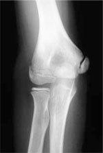
Little League Elbow (medial epicondylar apophysitis)

Little League Elbow (medial epicondylar apophysitis)

These injuries are just some of the most common ones I see.  In the article, the reporter focused on baseball and throwing.  With that in mind, consider research published in the American Journal of Sports Medicine this past February from renowned surgeon James Andrews that revealed players who pitch more than 100 innings in a calendar year are 3.5 times more likely to be injured.

He goes on to say that “these injuries are the result of a system that prepares genetically gifted athletes to play at the highest levels, but eliminates most players because their bodies cannot withstand such intense activity at such an early age.”  Sadly, he told the reporter that in 1998 he performed the Tommy John procedure on 5 kids high school age or younger, while in 2008 he did the same procedure on 28 children in the same age range.  This injury is usually caused by throwing too much too soon.

Consider the following data on suggested pitch counts per game (source James Andrews, MD & Glenn Fleisig, MD):

  • 8-10 y/o = 52 plus/minus 15 pitches
  • 11-12 y/o = 68 plus/minus 18
  • 13-14 y/o = 76 plus/minus 16
  • 15-16 y/o = 91 plus/minus 16
  • 17-18 y/o = 106 plus/minus 16

Unfortunately, I can personally relate to this blog post and story.  I was a promising young pitcher up until the point I threw my arm out in travel baseball at age 13.  The pain got so bad in my arm I could barely throw a ball 10 feet.  I remember the orthopedic surgeon telling me that I could not throw again the rest of the summer.  The pain (and memory of it) was so bad I elected to focus on position play and not to pitch again until my senior year of high school.  At that point, my arm was no longer the same as I had missed three years of practice and development.  Now, I too had become one of those kids whose body was never the same.

So, as a rehab and strength & conditioning professional, I want to help educate and promote better awareness to athletes, parents, coaches, trainers, AD’s, ATC’s and anyone who is involved in the care and training of young athletes.  Fortunately, people are taking positive steps to reduce overuse injuries.  One great initiative is STOP – Sports Trauma Overuse Prevention and you can learn more by clicking here to visit their website.

In the end, we must continue to educate everyone that the old motto of “No Pain, No Gain” is NOT the way to handle overuse injuries as this mentality may ruin the careers of young athletes or lead to an otherwise preventable injury and/or premature musculo-skeletal damage.  Pain truly is a warning signal the body gives us to detect mechanical problems and make changes in our training/activity level until we sort out the cause and solution.  I hope you will join me in supporting this mission and working hard at making sports fun, safe and free of overuse injuries for young athletes of all ages in the years to come.

References – The News & Observer – May 15, 2011